Case 4
A 56 year old patient consults our emergency department due to dysphagia which starting a couple of weeks ago.

Chest x-ray PA: complete, homogenes left lung opacity.
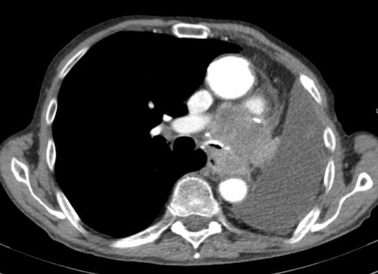
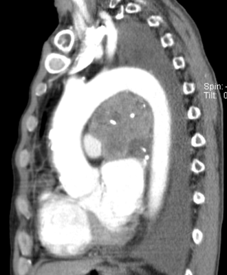
The patient received a resection of the left lung due to a bronchial carzinoma, almost exactly one year ago. In consequence, the chest x-ray shows the expected, complete opacification of the resected side. Further assessment of the dysphagia included a gastroscopy and endosonography with the following findings...
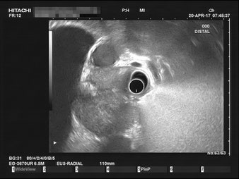


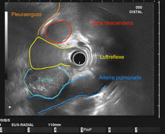

After an unremarkable gastroscopy, the endosonography revealed a large tumorous mass, spanning from the level of the middle esophagus to the heart. The imaging raises the suspicion that the mass is partly compresses the pulmonary artery. The tumor broadly infiltrates the esophagus, mostly likely the cause of the dysphagia. For a better anatomical overview, we have added the CT scans: