
Case 5
A 45 year old patient with known familial adenomatous polyposis syndrome (FAP) comes to his routine gastroscopy. The patient experienced a gradual weight loss in the last 3 months. The endoscopic inspection reveals flat erosions of the mucosal layer in the body of the stomach. EUS was preformed for further assessment.
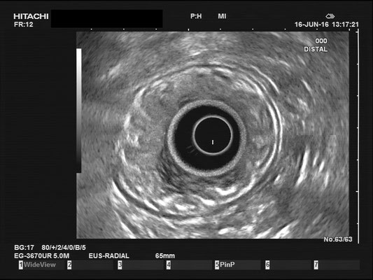
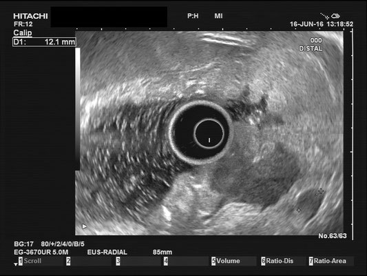


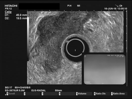
Contrary to the fairly localized erosion of the mucosal layer, the endosonographic image unmasked a large tumor expansion in the deeper layers of the mucous membrane. The hypoechoic tumor measured approximately 5cm, exceeding the muscularis propria.
Tumore und endosonographisches Staging
Eine der Limitationen der Endosonographie ist die korrekte Unterscheidung zwischen einem Tumorstadium T2 und T3. Wichtig? JA, weil die onkologische Behandlung in den Stadien zwischen einen primären OP (T2) und einer neo-adjuvanten Chemotherapie (also Chemo-OP-Chemo; T3) variiert.
Manche Experten empfehlen eine Unterteilung in Stadium T3 early entsprechend <2mm "Tumorinfiltration" und T3 advanced >2mm. Eventuell kann ein Patient mit T3 early so behandelt werden wie ein T2 Tumor und somit die neo-adjuvante Therapie erspart bleiben.
Lymphknoten und endosonographisches Staging
Die Genauigkeit der uN-Klassifikation mittels Endosonographie liegt bei ca. 65%. Genauer gesagt ist die Sensitivität für N1 94%, die Spezifität 20%, was bedeutet, dass ca. 80% der N0 Patienten "overstaged" sind.